The humerus is the long bone in the arm that runs from the shoulder to the elbow. It plays a crucial role in the movement and stability of the upper limb. Understanding the humerus bone markings is all-important for medical professionals, anatomists, and students of human anatomy. These markings function as attachment points for muscles, ligaments, and tendons, and they provide valuable info about the bone's part and construction.
Anatomy of the Humerus
The humerus is separate into several distinct regions, each with its own set of humerus bone markings. These regions include the head, neck, body, and distal end. The head of the humerus articulates with the glenoid cavity of the scapula to form the shoulder joint. The body, or shaft, of the humerus is the long, cylindrical portion that extends from the neck to the distal end. The distal end includes the sidelong and medial epicondyles, the trochlea, and the capitulum, which articulate with the bones of the forearm to form the elbow joint.
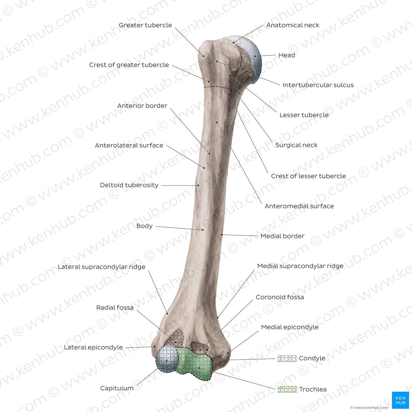
Proximal Humerus Bone Markings
The proximal end of the humerus features various important humerus bone markings that are crucial for understanding the bone s part and the muscles that attach to it. These markings include:
- Head of the Humerus: This is the rounded, smooth surface that articulates with the glenoid cavity of the scapula.
- Anatomical Neck: This is a slight constriction just below the head of the humerus.
- Greater Tubercle: This is a large, rounded swelling on the lateral side of the humerus, which serves as an attachment site for the rotator cuff muscles.
- Lesser Tubercle: This is a smaller prominence on the median side of the humerus, which also serves as an attachment site for the rotator cuff muscles.
- Intertubercular Groove: This is a deep groove that runs between the greater and lesser tubercles, providing a passage for the tendon of the long head of the biceps brachii muscle.
Shaft of the Humerus
The shaft, or body, of the humerus is comparatively smooth and cylindrical, with a few noteworthy humerus bone markings. These include:
- Deltoid Tuberosity: This is a rough, V shaped area on the sidelong side of the shaft, which serves as an attachment site for the deltoid muscle.
- Radial Groove: This is a shallow groove on the tail surface of the shaft, which provides a passage for the radial nerve and the profunda brachii artery.
- Nutrient Foramen: This is a small open on the anterior surface of the shaft, through which blood vessels enter the bone to supply it with nutrients.
Distal Humerus Bone Markings
The distal end of the humerus features respective crucial humerus bone markings that are important for translate the bone s use and the muscles that attach to it. These markings include:
- Lateral Epicondyle: This is a spectacular bony process on the sidelong side of the distal humerus, which serves as an attachment site for the extensor muscles of the forearm.
- Medial Epicondyle: This is a prominent bony procedure on the medial side of the distal humerus, which serves as an attachment site for the flexor muscles of the forearm.
- Trochlea: This is a smooth, pulley forge surface on the medial side of the distal humerus, which articulates with the trochlear notch of the ulna to form the elbow joint.
- Capitulum: This is a smooth, round surface on the sidelong side of the distal humerus, which articulates with the head of the radius to form the elbow joint.
- Coronoid Fossa: This is a shallow slump on the anterior surface of the distal humerus, which accommodates the coronoid summons of the ulna during flexion of the elbow.
- Olecranon Fossa: This is a deep slump on the bum surface of the distal humerus, which accommodates the olecranon summons of the ulna during extension of the elbow.
Clinical Significance of Humerus Bone Markings
Understanding the humerus bone markings is crucial for diagnosing and treating various injuries and conditions affecting the humerus. for instance:
- Fractures: Fractures of the humerus can occur at assorted points along the bone, and cognition of the humerus bone markings can facilitate identify the specific position and type of fracture.
- Dislocations: Dislocations of the shoulder or elbow joint can cause damage to the humerus bone markings, and understanding these markings can aid in diagnosing and treat these injuries.
- Muscle and Tendon Injuries: Injuries to the muscles and tendons that attach to the humerus bone markings can have pain and limited range of motion. Knowledge of these markings can help identify the specific muscles or tendons involved and guide treatment.
Note: The humerus bone markings are also important for operative procedures involve the humerus, such as joint replacements or fault repairs. Surgeons must have a thorough read of these markings to ensure proper placement of implants and to avoid damaging nearby structures.
Imaging Techniques for Visualizing Humerus Bone Markings
Several visualize techniques can be used to visualize the humerus bone markings and diagnose injuries or conditions regard the humerus. These techniques include:
- X rays: X rays are commonly used to envision the humerus and its humerus bone markings. They can assist identify fractures, dislocations, and other abnormalities.
- Computed Tomography (CT) Scans: CT scans provide detailed cross sectional images of the humerus and its humerus bone markings. They are useful for name complex fractures and planning surgical procedures.
- Magnetic Resonance Imaging (MRI): MRI scans render detail images of the soft tissues ring the humerus, as well as the bone itself. They are utile for diagnose muscle and tendon injuries, as well as other conditions affecting the humerus bone markings.
Common Injuries and Conditions Affecting the Humerus
Several injuries and conditions can affect the humerus and its humerus bone markings. Some of the most common include:
- Fractures: Fractures of the humerus can occur at diverse points along the bone, include the proximal, shaft, and distal regions. Common types of humerus fractures include:
| Type of Fracture | Description |
|---|---|
| Proximal Humerus Fracture | A cracking that occurs near the head of the humerus, often involving the greater or lesser tubercles. |
| Humeral Shaft Fracture | A cracking that occurs along the shaft of the humerus, often have by unmediated trauma or a fall. |
| Distal Humerus Fracture | A fracture that occurs near the distal end of the humerus, oftentimes involving the lateral or median epicondyles. |
- Dislocations: Dislocations of the shoulder or elbow joint can get damage to the humerus bone markings and surrounding tissues. Common types of dislocations include:
| Type of Dislocation | Description |
|---|---|
| Shoulder Dislocation | A dislocation that occurs when the head of the humerus is force out of the glenoid caries of the scapula. |
| Elbow Dislocation | A dislocation that occurs when the distal end of the humerus is force out of alignment with the bones of the forearm. |
- Muscle and Tendon Injuries: Injuries to the muscles and tendons that attach to the humerus bone markings can make pain and determine range of motion. Common types of muscle and tendon injuries include:
| Type of Injury | Description |
|---|---|
| Rotator Cuff Tear | An injury that occurs when one or more of the rotator cuff tendons are torn, often regard the greater or lesser tubercles of the humerus. |
| Biceps Tendonitis | An injury that occurs when the tendon of the long head of the biceps brachii muscle becomes inflamed, often imply the intertubercular groove of the humerus. |
Note: Treatment for injuries and conditions affecting the humerus and its humerus bone markings may include rest, ice, compaction, elevation (RICE), physical therapy, medicament, or surgery, depending on the asperity of the injury.
Conclusion
The humerus is a complex bone with legion humerus bone markings that play crucial roles in the movement and constancy of the speed limb. Understanding these markings is essential for name and treating assorted injuries and conditions regard the humerus. By acquaint themselves with the anatomy and clinical significance of the humerus bone markings, aesculapian professionals, anatomists, and students can gain a deeper appreciation for the function and construction of this crucial bone.
Related Terms:
- humerus bone markings quiz
- femur bone markings
- humerus bone markings chart
- radius bone markings
- humerus bone unlabeled
- humerus bone markings labeled